
BIDS-Processed Data Survey Results
Introduction
We launched a survey to investigate what derivatives the neuroimaging community determines has a strong chance of being reused or has a higher priority to save and share. The survey was shared across several prevalent neuroimaging mailing lists. These results will guide our standardization efforts. The survey focused on derivatives for MRI modalities – anatomical, functional, and diffusion imaging. We decided the derivative options should be software agnostic to better gauge the outputs the community determined are more generally necessary for reusability and sharing rather than for particular software or analysis. We had a rating system that went from: must have, useful, likely will never look at, and don’t know what this is. We received 139 responses! The responses can be found here. In this blog post we will report some of the results found from each modality.
Table of Contents
- MRI Anatomical Imaging results
- MRI Functional Imaging results
- MRI Diffusion Imaging results
- General templates and quality control
- Discussion
1.MRI Anatomical Imaging results
For MRI anatomical imaging, we found several outputs reached over 50% of the total responses marked as either ‘must have’ or ‘useful’. The outputs with over 50% of responses marked ‘must have’ were: registered image in a template space (i.e. MNI), final preprocessed output image from pipeline (i.e. N4 corrected and merged T1w image for multiple T1w’s), skull stripped brain image, and the transformation file to MNI space (i.e. .mat or .h5). The outputs with over 50% of responses marked ‘useful’ were: other structural modalities (i.e. PD, T2w, etc) registered to T1w, cortical surfaces, and the bias corrected image. We have illustrated below the percent of total responses for the ranking of ‘must have’ or ‘useful’ across all the outputs (Figure 1 and Figure 2 respectively).

Figure 1. MRI anatomical imaging ‘must have’ responses for all outputs ordered greatest to least.
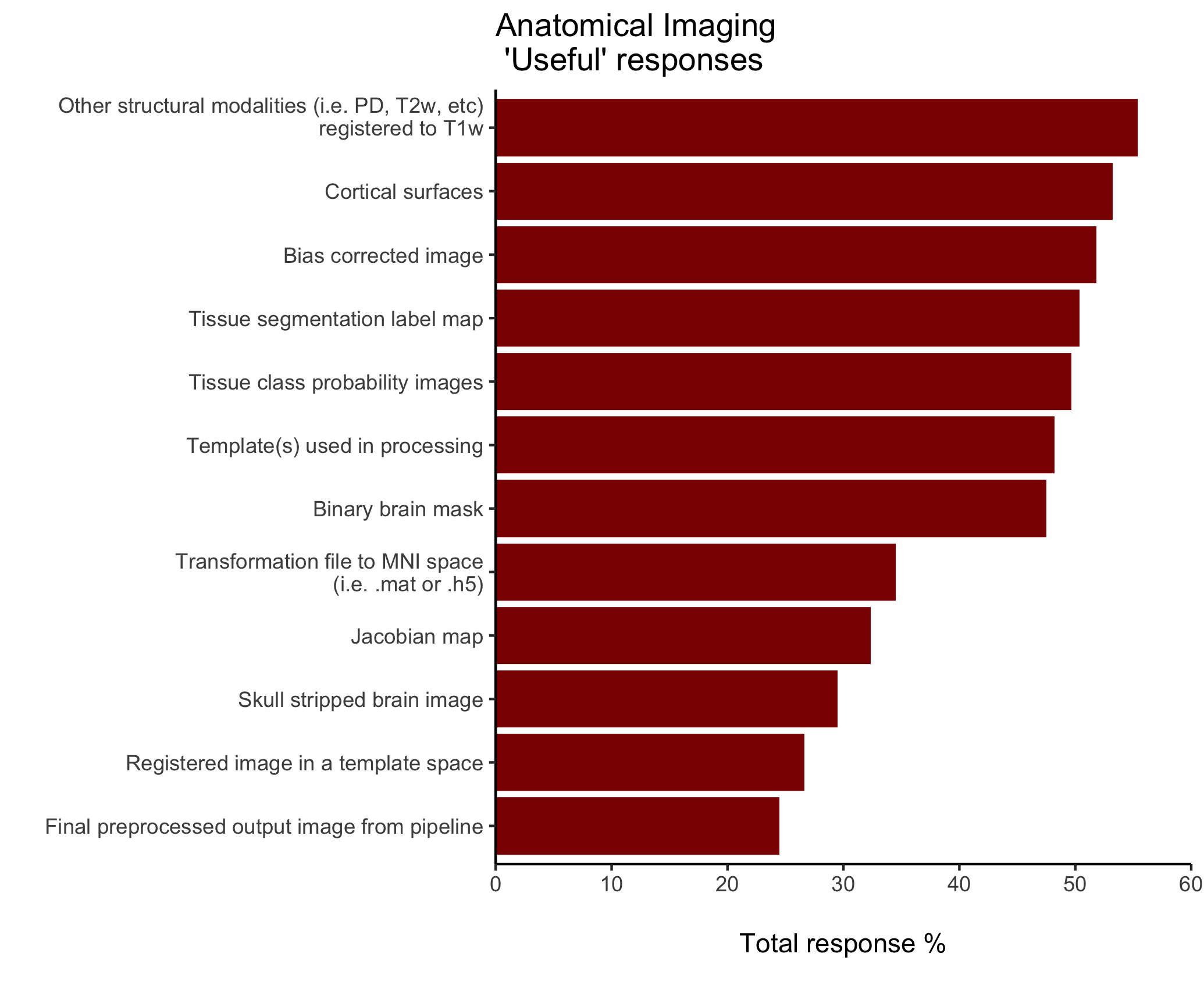 Figure 2. MRI anatomical imaging ‘useful’ responses for all outputs ordered greatest to least.
One derivative that received the highest marks for both ‘likely will never look at’ and ‘don’t know what this is’ was the jacobian map. The additional outputs were more familiar to respondents and received no more than 6% of the ‘don’t know what this is’ option.
Valuable anatomical outputs that weren’t captured in the ranking evaluation and commonly recommended in the free response section were: lesion segmentation masks and quality control results.
2. MRI Functional Imaging results
For MRI functional imaging, we found several derivatives achieved over 50% responses marked with the highest rating (‘must have’). The outputs that satisfied this metric were: final preprocessed output from pipeline (i.e. BOLD image corrected and sampled to template or output space), registered output in a template space (i.e. MNI), confound regressors, transformation file to T1w space (i.e. .mat or .h5). We have displayed below the percent of total responses for the ranking of ‘must have’ across all outputs (Figure 3).
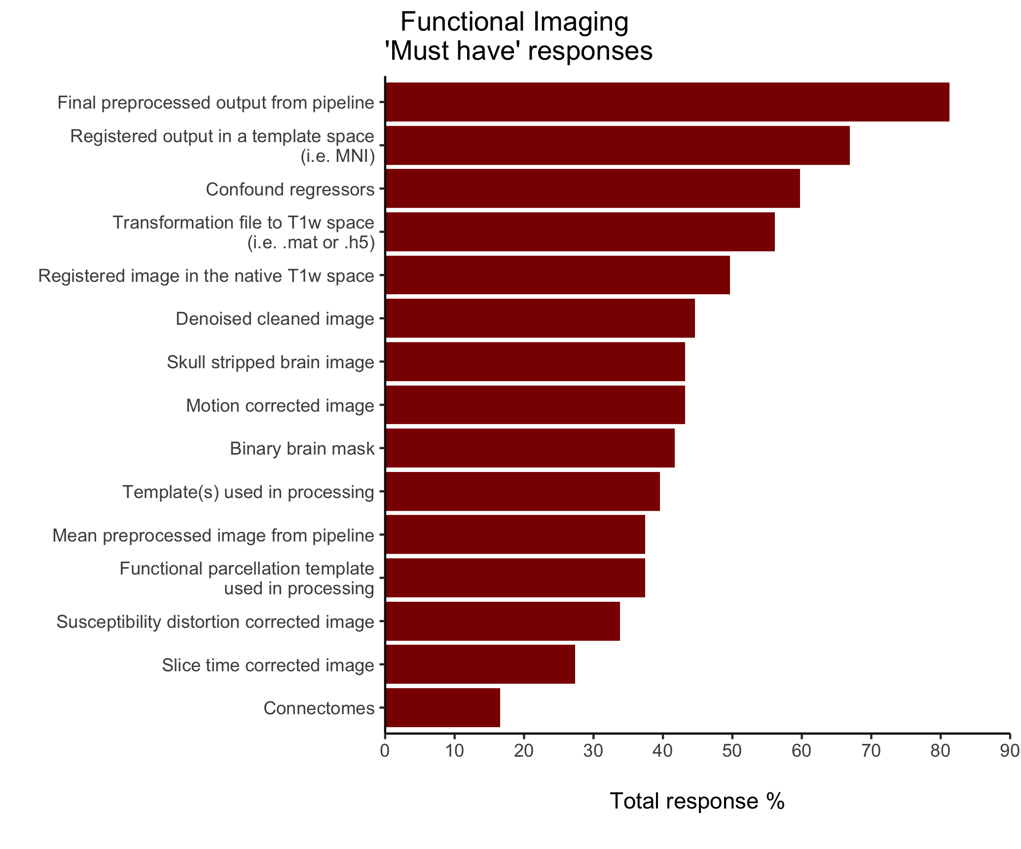
Figure 3. MRI functional imaging ‘must have’ responses for all outputs ordered greatest to least.
We found only one derivative, connectomes, had over 50% of responses marked with the second strongest rating (‘useful’). The respondents were more familiar with functional outputs due to the ‘don’t know what this is’ option across all the outputs received less than 6% of responses. Some valuable functional outputs that were not captured in the ranking evaluation and commonly recommended in the free response section were: ICA components and quality control results.
In addition, we were interested in eliciting feedback regarding cortical analysis. We found that about 51% of our respondents run analysis on cortical surfaces. From those that run analysis on cortical surfaces they prefer to target the individual subject (~110k vertices) and 32k_fs_LR (Human Connectome Project). These were the only options that had over 45% of the responses.
3. MRI Diffusion Imaging results
For MRI diffusion imaging, we found on average about 25% of our respondents marked ‘don’t know what this is’ across the outputs. The largest ‘don’t know what this is’ option was for the tractogram file. Interestingly, for all outputs the ‘likely will never look at’ ranking received 15% or less of the responses. Thus, a majority of the responses indicate that either the outputs identified are ‘must have’, ‘useful’, or ‘don’t know what this is’. This made it intriguing to evaluate the difference between ‘must have’ and ‘useful’ responses. The outputs with more pronounced difference (greater than ±15%) were: model-derived maps (i.e. FA, MD, AD, RD), eddy corrected image, and deterministic tractography image. We have pictured below the difference between these responses across all the outputs (Figure 4).
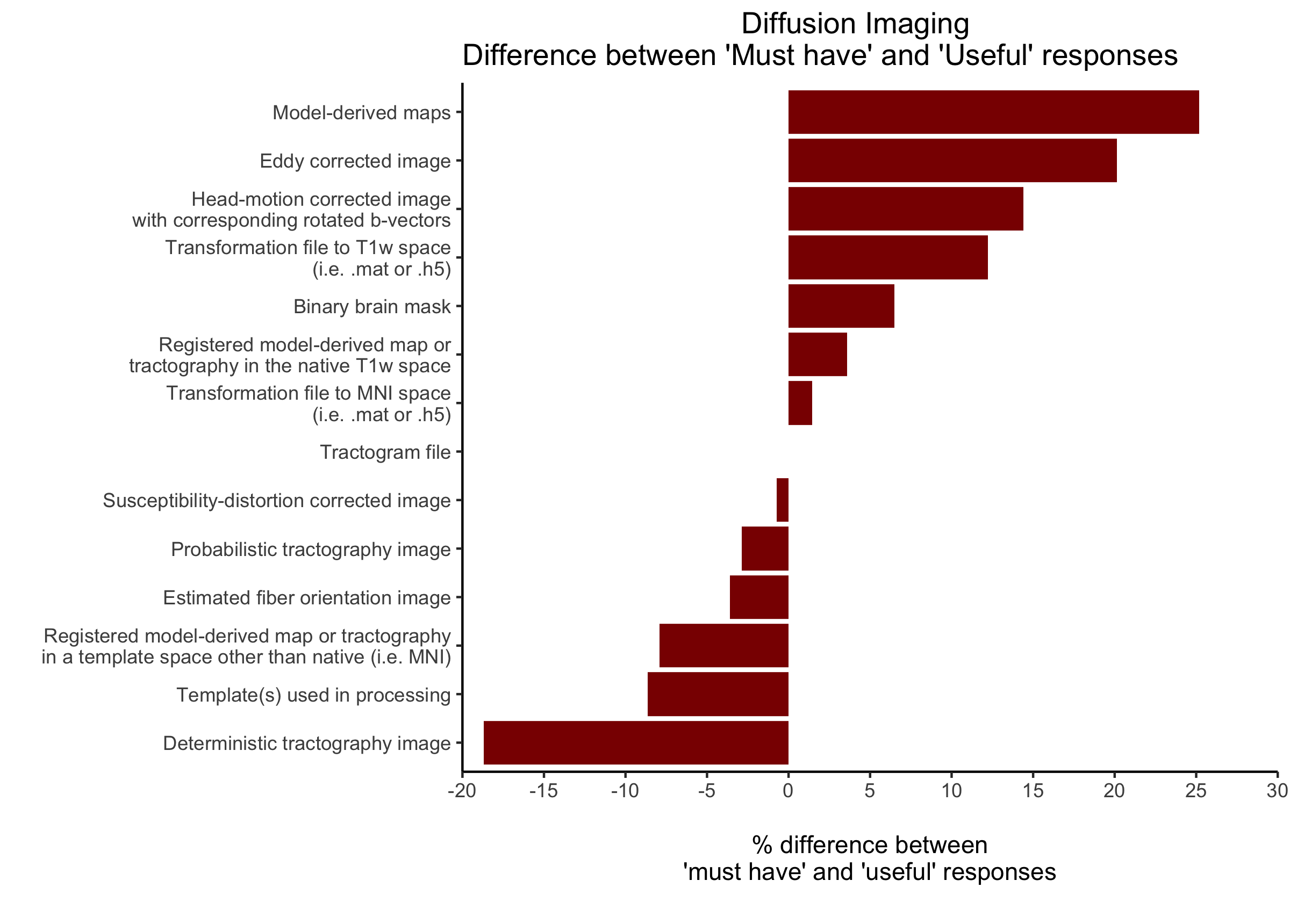
Figure 4. MRI diffusion imaging difference between ‘must have’ and ‘useful’ responses for all outputs ordered greatest to least. A positive value indicates the ‘must have’ is greater than ‘useful’ response. A negative value represents the ‘useful’ is greater than ‘must have’ marking.
Valuable diffusion outputs commonly suggested in the free response area were: TBSS skeleton and quality control results.
4. General templates and quality control
We were interested in what preferred template space and file formats data was in and quality control approaches the community performed. We found the community prefer their template space to be in MNI or fsaverage. In addition, the file formats preference was nifti or gifti. The typical quality control strategies implemented use either a visual approach or quantitative summaries. For the visual approach, we found the common ways that respondents visualized to quality control were through fsleyes (or fslview), freeview, an html page, or another brain viewer software to evaluate the analysis quality (i.e. segmentation or registration). We found for the quantitative reports responses generally utilized the reports provided by running MRIQC, eddyqc, DVARS stats, evaluated motion parameters, assessed the confounds, or another summary report.
This survey has provided us valuable insights into what the different derivatives the neuroimaging community determines may be necessary for data reusability and sharing. These insights will provide us with an idea of how to focus our standardization efforts within the processed data domain. In both anatomical and functional imaging derivatives two outputs received over 50% of the strongest marking (‘must have’): final preprocessed output image from a pipeline and the transformation file to another space (i.e. .mat or .h5). We found a free response comment that persisted across the modalities was the need to share the quality assessment results, metrics, or logs used to record or make the decision on the processed image quality. The diffusion imaging derivatives could be sampled more due to a quarter of the current respondents don’t know or don’t do diffusion imaging. The results for anatomical and functional imaging are perhaps more robust due to less than 6% of respondents didn’t know what the output was.
We were interested in determining what outputs across modalities the current BIDS extension proposals may already cover. For the MRI anatomical imaging outputs with over 50% of ‘must have’ markings we found the final preprocessed output image from a pipeline, registered image in a template space, and the skull stripped brain image is described in the BIDS Extension Proposal (BEP) 3 and the transformation file (i.e. .mat or .h5) is covered in BEP 14. The anatomical outputs with over 50% of ‘useful’ markings described in an extension are cortical surfaces in BEP 11 and other anatomical modalities registered to T1w and the bias corrected image are in BEP 3. In MRI functional imaging outputs with over 50% of ‘must have’ marking, we found the final preprocessed output from a pipeline and the registered output in a template space are described in BEP 3, confound regressors are described in BEP 12, and the transformation file to T1w space is covered in BEP 14. Connectomes received over 50% of responses for ‘useful’ and can be found in BEP 17. The MRI diffusion imaging outputs with the most pronounced difference between ‘must have’ and ‘useful’ responses covered are the model-derived maps and deterministic tractography image in BEP 16 and the eddy corrected image in BEP 3.
We thank the neuroimaging community for their time and valuable feedback!

Submit a comment